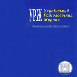
UJR 2004, vol XII, # 1

THE CONTENTS
2004, vol 12, № 1, page 7
O. P. Sharmazanova, D. А. Mitelyov
Clinical x-ray changes of cervical spine in connective tissue systemic dysplasia in teen-agers
Annotation
Objective: To study vertebrological signs of connective tissue systemic dysplasia (CTSD) at the level of cervical spine in teenagers using clinical and x-ray findings.
Material and Methods: Standard x-ray pictures and functional spondylograms of the cervical spine (CS) were studied visually and using radiogrammetry in 38 boys aged 13-17 in non-differentiated CTSD.
Results: It was determined that x-ray changes at the level of CS were observed in all patients. Local and systemic anomalies were revealed in 42.1 % , instability in 68.4 % , deforming spondyloarthrosis in 63.1 % of the patients. In 39.4 % of cases primary manifestations of osteochondrosis were noted. The clinical picture was characterized by the changes in the nervous and musculoskeletal systems.
Conclusion: In CTSD cervical spine is frequently involved therefore further studies of this pathology are necessary with the consideration of the possibility of the disease progress.
Key words: connective tissue systemic dysplasia, teen-agers, cervical spine, x-ray studies.
2004, vol 12, № 1, page 11
V. I. Grishenko, O. V. Grishenko, O. V. Mercalova, O. Y. Geleznyakov
Comparison of MRI capabilities in ovarian benign tumors visualization
Annotation
Objective: To improve the quality of diagnosis of ovarian benign tumors and tumor-like formations by working out optimal methodological approaches to MRI performing in this pathology.
Material and Methods: One hundred and twenty four women with ovarian pathology and 17 healthy women were examined. MRI of the organs of the small pelvis was performed with a 0.12 T MRI unit "Obraz-1" (Russia). Examinations were carried out in T1 and T2 weighted modes. Quantitative (size) and qualitative (localization, form) parameters of MR images were analyzed. Statistical analysis of the data obtained was made by "Medstat" database.
Results: A high diagnostic significance of MRI in examinations of women with ovarian neoplasms was demonstrated. The most optimal MRI modes for internal genitalia were defined, that allowed us both to differentiate benign ovarian tumors and tumor-like formations and to assess the morphological structures of ovarian tumors.
Conclusions: MRI has a crucial importance for a differential diagnosis of the ovarian benign tumors and tumor-like formations. The use of MRI parameters such as relaxation time of proton density, signal intensity allow us both to diagnose ovarian pathology and to suppose a morphological structure of voluminous ovarian formations.
Key words: MRI, ovarian benign tumors, tissue parameters of tumors.
2004, vol 12, № 1, page 14
R. Brendene, V. Mamontovas
Radiodiagnosis of mediastinum invasion by lung cancer
Annotation
Objective: To analyse correlation between lung cancer morphology, growth type, localization and mediastinal invasion.
Material and Methods: The data of 372 patients were analysed. Mediastinal invasion ( ТЗ , Т 4) was histologicaly confirmed in 128 (34.4 %) patients. Aorta invasion was confirmed in 21 (5.6 %), superior vena cava in 11 (2.9 %), pericardium in 55 (14.8 %), esophagus in 7 (1.9 %) patients. Chest radiography was performed in all patients; 68 patients underwent chest CT.
Results: It was estimated that probability of mediastinal invasion increased in case of squamous cell carcinoma (p<0.05). At marked criteria of possible mediastinal invasion - 3cm or more mass contact with mediastinum, more than 90 degrees of contact with aorta, obliteration of the fat between the mass and mediastinal structures — sensitivity of CT increased in identifying mediastinal invasion and operability from 33 % to 57 %.
Conclusions: Mediastinal invasion depends on the morphology of cancer. Radiographic signs such as mass contact with mediastinum, blurring of aortic arch, mediastinal pleura thickening may preclude mediastinal invasion. CT criteria of possible mediastinal invasion may suggest the process operability.
Key words: lung cancer, histological forms, mediastinal invasion, operability.
2004, vol 12, № 1, page 19
S. V. Afanasev
Radioisotope study of pancreatic external secretion and intestinal absorption
Annotation
Objective: To work out a physiological, cost-effective, accessible, specific, and informative technique for radioisotope study of pancreatic external secretion and intestinal absorption.
Material and Methods: The study involved 10 healthy persons and 29 patients with chronic pancreatitis. External secretion of the pancreas and intestinal absorption was investigated using the technique for radioisotope diagnosis worked out in the hospital of Ukrainian State Research Institute for Medical and Social Problems of Handicapped Persons.
A piece of hard animal fat (1 cm3) was used as a fat carrier, technetium 99m — as a radioactive tracer. The study was performed using gamma-camera GSK-301-T, the information was stored on a 64x64 matrix. The findings were registered in the real-time mode.
Results: The obtained findings allowed to determine time intervals of the fat carrier lipolysis, absorption and entrance of the isotope to the urinary bladder in persons without disturbances of pancreatic external secretion and intestinal absorption as well as in patients with chronic pancreatitis with different degree of severity of the above disturbances.
Conclusion: The developed technique for radioisotope study of pancreatic external secretion and intestinal absorption with the use of hard animal fat as a carrier is physiological, cost-effective, specific and informative, it can be widely used in Ukraine . The use of the described technique in clinical practice and expertise allows to improve the quality and efficiency of diagnosis and treatment of the patients with chronic pancreatitis.
Key words: pancreas, intestine, external secretion, absorption, radioisotope study.
2004, vol 12, № 1, page 23
V. М. Slavnov, S. V. Bolgarskay, Е. V. Taran, V. V. Markov
Influence of antibacterial therapy on bone scan indices at foot inflammation in diabetes mellitus accompanied by diabetic foot syndrome
Annotation
Objective: To study the influence of antibacterial therapy on bone scan indices at foot inflammation in patients with diabetes mellitus (DM) accompanied by diabetic foot syndrome.
Material and Methods: The original technique was used to examine 29 patients with severe type 2 DM in decompensation stage accompanied by diabetic foot syndrome with trophic ulcers, of them 17 patients before treatment and 12 patients after antibacterial therapy. Bone scan was performed using scintillation tomographic gamma-camera ГКС 301 T3 hours after intravenous injection of 600 MBq of 99mTc-methylene diphosphonate.
Results: Considerable accumulation of the RP in the lesion focus both in the anterior and median foot portions was observed in DM patients with diabetic foot syndrome and trophic ulcers of the lower extremity at bone scan. The majority of the patients did not have x-ray signs of inflammatory process in the bones of the anterior portion of the foot. Antibacterial therapy of DM patients included the use of osteotropic antibiotics and metronidazole. The treatment was administered for 8-12 weeks. After the course of treatment, significant reduction in summary activity in the foot and percentage of asymmetry of summary activity between the feet were noted.
Conclusion: DM patients with diabetic foot syndrome presented with purulent inflammation in the bones of the anterior foot. Prolonged antibacterial therapy was accompanied by resolution of the trophic ulcers and transition of the acute inflammatory process to remission phase of chronic osteomyelitis which was confirmed by the radionuclide studies.
Key words: bone scan, gamma-camera, 99mTc-methylen diphosphonate, diabetes mellitus, diabetic foot, osteomyelitis.
2004, vol 12, № 1, page 26
А. V. Svinarenko
Chronomodulation of concomitant radiochemotherapy for primary inoperable rectal cancer
Annotation
Objective: To assess immediate results of treatment for primary inoperable rectal cancer (RC) using a new protocol of treatment which consists in simultaneous administration of radiation and chemotherapy in chronomodualted mode.
Material and Methods: Main group consisted of 10 patients with stage 2 RC (T3-4N0M0) and stage 3 RC (T3 - 4N1M0) aged 25—67 (6 men and 4 women) who received distance gamma-therapy with medium fractions (4Gy twice a week) until 32 Gy MFD at small pelvis. Each radiotherapy treatment was preceded by bolus administration of leucovorin (20mg/m2) followed by 12-hour IV infusion of 5-fluoruracil (500mg/m2) which terminated 6—8 hours before the irradiation. 3-4 weeks after chronomodulated radio-chemotherapy (CRCT) operability of the tumor was assessed and the surgery was performed. The controls were 10 patients who were treated using a generally accepted protocol (40 Gy with traditional fractions).
Results: CRCT was completed in 9 of the 10 patients. All the 9 patients were operated. In the controls only 6 tumors were made resectable. The original CRCT protocol allows to reach resumption of the primary tumor and increase the degree of radiation changes in it. We think that this factor guarantees higher percentage of resectable tumors.
CRCT was better tolerated than the generally accepted protocol. Only in one case CRCT was terminated due to the disease progress and stage 3 toxic effects (nausea, diarrhea, leucopenia). In the rest 9 patients the course of treatment was not intermitted, toxic reactions did not exceed I-II degree. In the controls the treatment was done with 6-10 days intervals in 4 patients due to cystitis, diarrhea, epidermitis and III degree leucopenia.
Conclusion: The use of CRCT allows to make RC resectable more frequently than at traditional treatment. The number and degree of toxic reactions at CRCT does not increase when compared with radiation therapy which is performed without the consideration of the time of the day.
Key words: rectal cancer, radiochemotherapy, chronomodulation.
2004, vol 12, № 1, page 31
N. А. Mitrayeva, М. А.Ishanova, V. P. Starenkiy, Т. S. Bakay, L. І. Grebenik
Evaluation of tumor marKer expression at radiotherapy efficacy monitoring in cancer patients
Annotation
Objective: To develop criteria of application of tumor markers (prostatic specific antigen (PSA, bombesin) for monitoring radiotherapy efficacy in prostate, breast and lung cancer.
Material and Methods: The study involved 82 patients with prostate cancer (PC), 80 patients with breast cancer ( ВС ) and 49 patients with lung cancer (LC). All patients were examined before the treatment and after radiotherapy course. Free to total PSA ration was measured in PC patients using IEA commercial kits (Microwell, USA).
Ca-15-3 and Ca-125 in ВС patients were evaluated using IEA kits (Diatec, USA). To study bombesin levels in the blood of LC patients commercially available 125I-RIA-kit ( DRY , USA ) were used.
Results: 95 % sensitivity and 80% specif ity for the ratio free to total PSA in PC patients (PSAtree/PSAtotal xl00%) was shown. It was established for PC patients that discriminative level of this ratio was equal to 12 %. Bombesin allowed to perform monitoring of radiotherapy efficacy in small-cell LC patients (traditional and accelerated fractionation) and in squamous cell LC patients (accelerated fractionation). Low informativity of using Ca-125 and Ca 15-3 tumor markers in ВС patients because of a low correlation between the results of immunoassay and clinical dates was shown.
Conclusion: The possibility to use PSA for monitoring of radiotherapy efficacy in prostate cancer was studied. Low efficacy of generally accepted tumor markers of breast cancer in evaluation of radiotherapy efficacy was proved to be due to low correlation of the results with clinical data. The possibility to use bombesin for monitoring of the efficacy of various irradiation modes in lung cancer was established.
Key words: radiation therapy, tumor markers, monitoring.
2004, vol 12, № 1, page 36
L. P. Abramova, L. І. Simonova, V. V. Myasoedov,
М. K. Adeyshvili-Siromyatnikova, S. M. Pushkar, O. І. Paskevich
Influence of radiation therapy on antioxidant homeostasis in patients with breast cancer
Annotation
Objective: To study the influence of radiation therapy on the state of lipid peroxidation and activity of main antioxidant enzymes in patients with breast malignancy.
Material and Methods: The state of lipid peroxidation (LP) was studied in patients with breast cancer ( ВС ) undergoing radiation therapy according to the amount of the following lipid peroxidation products: dien conjugates and malonic dialdehyde. The degree of antioxidant ( АО ) protection in these patients was evaluated according to the activity of antioxidant enzymes: superoxide dysmutase, catalase, glutathione peroxidase, ceruloplasmin.
Results: Primary examination of the patients with ВС revealed strained condition of lipid peroxidation which was accompanied by disbalance of enzyme systems of АО protection. Antitumor treatment with the use of high doses of ionizing radiation (mean focal dose 40 Gy) caused constant induction of free radicals and development of chain reactions of lipid peroxidation, which resulted in extreme use of stress-limiting АО systems due to the necessity of constant blockage of peroxidation processes and in turn increased the amount of LP derivates at АО potential exhaustion.
Stabilizing of LP due to increased consumption of stress-limiting АО systems, especially glutathione peroxidase and vitamins С and E was observed after radical surgery in ВС patients. The use of radiation therapy caused secondary increase in peroxide processes due to constant induction of free-radical oxidation and abundant use of АО protection.
Conclusion: The obtained results suggest the necessity of administration of exogenic АО drugs in complex treatment which accompanies radiotherapy in cancer patients.
Key words: breast cancer, radiation therapy, lipid peroxidation, dien conjugates, malonic dialdehyde, antioxidant enzymes, catalase, superoxide dysmutase, glutathione peroxidase, ceruloplasmin.
2004, vol 12, № 1, page 40
G. А. Zamotaeva, N. М. Stepura, N. V. Sologub
Effect of thymic preparations on thymulin level and circulating immune complexes at experimental iodine-131 incorporation
Annotation
Objective: Experimental study of the effect of different doses of iodine-131 on the content of thymulin and circulating immune complexes, and possibility to use thymus preparations with the purpose of immunorehabilitation.
Material and Methods: This work was carried out on male CBA mice. 1-131 was injected intraperitoneally at a dose of 0.925 and 9.25 kBq/g. Tactivin was administered subcutaneously at a dose of 1 meg, thymogen was injected intramuscularly at a dose of 0.1 meg every other day — a total of 10 injections. The content of thyroxine, triiodothyronine, circulating immune complexes (CIC) and thymulin was determined. The studies were performed on days 7,14, 28, 56, and 84 after isotope administration.
Results: It was established that single 131 I parenteral administration at a dose of 0.925 and 9.25 kBq/g (absorbed doses were 5.38 and 56 Gy, respectively) caused dose-dependent changes in the immune status of the mice: increase in CIC level and inhibition of thymic endocrine function. The use of immunomodulators (tactivin and thymogen) in animals exposed to radioiodine effect at a dose of 9.25 kBq/g stimulated thymic hormone production and decreased CIC level.
Conclusion: Iodine-131 incorporation at a dose of 0.925 and 9.25 kBq/g caused a significant increase in CIC level and inhibition of thymic endocrine function, which manifested by changes in blood serum thymulin content. Intensity, degree, and duration of immunologic changes depended on the dose of injected isotope and thyroid hormone level. Immunomodulators (thymogen and tactivin) may be effectively used for immunorehabilitation at 131 I incorporation.
Key words: Iodine-131, thyroid hormones, thymulin, circulating immune complexes, immunocorrection, thymogen, tactivin.
2004, vol 12, № 1, page 45
М. O. Klimenko, М. І. Onishenko
Influence of low-energy gamma-radiation on rat blood serum chemiluminescence at chronic inflammation
Annotation
Objective: To study dose dependence of chemiluminescence (CL) intensity at exposure to low-intensity gamma-radiation against a background of chronic inflammation.
Material and Methods: The study involved 96 Wistar male rats. Chronic inflammation was caused by injection of 4 ml 2 % caraginen solution to a preliminary prepared skin sac. Irradiation was delivered on day 3 and 7 of inflammation at a dose of 0.1, 0.5, 1.0 Gy (dose rate 20 mGy/h). Peak and residual intensity of blood serum chemiluminescence was assessed after addition of 2 ml of 10 % hydrogen peroxide to the sample.
Results: By day 3 of inflammation, the irradiation caused increase in maximum intensity of blood serum CL, the effect was observed 4 days after the irradiation (day 7 of inflammation). Linear dependence of the effect on the dose was noted. By day 7 the irradiation caused changes in maximum CL intensity only at a dose of 1.0 Gy, immediately after the irradiation this value increased and 7 days later (day 14 of inflammation) it decreased.
Conclusion: Our findings suggest the possibility of DNA damage by the products of lipid peroxidation at the mentioned doses and terms of inflammation as well as increase of oncogenic potential of chronic inflammation.
Key words: chronic inflammation, low-intensity gamma-radiation, chemilumenescense, lipid peroxidation.
2004, vol 12, № 1, page 49
O. М. Kovalenko, D. O. Biliy
Physical worKing ability in persons who survived acute radiation sicKness due to Chornoby accident (the data of 16-year follow-up)
Annotation
Objective: To study physical working ability (PWA) in male patients who survived acute radiation sickness (ARS) due to the accident at Chornobyl Atomic Power Plant during the years after the accident.
Material and Methods: PWA was studied in 30 persons who survived stage 1 ARS, 31 - stage 2 ARS, 5 - stage 3 ARS and 59 patients with absorbed doses of radiation < 1 Sv who did not demonstrate bone-marrow syndrome. The findings of longitudinal study were divided into 4 time periods: 1987-1988, 1989-1991, 1992-1996, 1997-2002. The controls were 20 healthy male subjects not exposed to radiation. The mean age of the patients from each group was similar. The study was performed using standard veloergometry with electrocardiography. The findings were processes using SPSS computer program.
Results: During the first 1-2 years after the accident PWA level in the exposed patients of all groups was considerably lower than in the controls and did not depend on the severity of the radiation exposure. It also did not considerably change later. The efficacy of hemodynamic provision of physical load (PL) in the patients and in the controls was similar. A number of types of reaction of the circulatory system to PL was distinguished: dystonic, hypertonic, ischemic, vague. Their incidence changed with the term of observation due to reduction in diastolic and increase in hypertonic types.
Conclusion: Considerable reduction in PWA in persons who survived ARS in the early period is chiefly caused by negative influence of ionizing radiation and is not determined by the degree of severity of bone-marrow syndrome. Further restoration of PWA does not occur which can be explained by development of neuro-somatic pathology against a background of age-related changes in the organs and systems of the victims.
Key words: physical working ability, ionizing radiation.
2004, vol 12, № 1, page 53
O. А. Romanova
The role of lymphoid pool of the cells in reconstitution of hemopoiesis of irradiated recipients of lymphomyelografts
Annotation
Objective: To study the dynamics of the reconstitution of lymphomyelopoiesis in irradiated recipients following syngenic combined lymphomyelotransplant injection.
Material and Methods: (CBAxC 57 BL)F 1 mice were irradiated at a dose of 9 Gy. Syngenic bone marrow (10x10 6 cells per mouse) and syngenic thymocytes (20 x 10 6 cells per mouse) were injected intravenously during the first 6-8 hours after irradiation. The animals were divided into the following groups: 1) irradiated animals, injected with bone marrow cells; 2) irradiated animals, injected with bone marrow cells and thymocytes; 3) normal nonirradiated animals.
The nature of alterations in the bone marrow cell pool of irradiation recipients was assessed by the dynamics of accumulation of colonyforming units (CFUs) in the femur, type of the formed colonies and their proportion, production of lymphocytes in the bone marrow, rate of accumulation of lymphocytes, their pheno-typical composition.
Results: The investigations showed that injection of syngenic thymocytes in combination with myelokaryocytes to the lethally irradiated recipients accelerated the compensation of the organism deficiency in hemopoietic units and in their successors. Lymphocytes accumulation rates in the bone marrow were more active in the recipients of the lymphomyelotransplant when compared to those of the myelotransplant. Index s m + / c m + s m - of cells ratio above one, characteristic for lymphopoiesis of normal mature animals, restored of lymphomyelotransplant recipients by the 30th day, and in recipients of myelotransplant — by the 45th day.
Conclisuon: The finding obtained in the experiments strongly suggest that addition of lymphoid cells to the bone marrow transplant enhances the processes maturation of in the colonyforming units in the bone marrow, their proliferation and differentiation in the irradiated recipients.
Key words: irradiation, lymphomyelotransplantation, hemopoiesis, lymphocytes.
2004, vol 12, № 1, page 58
Е. O. Romodanova, Т. S. Dyubko, Т. F. Morozova, V. O. Timanyuk
Spectral characteristics of BSA aqueous solutions at low-energy laser irradiation of solution components
Annotation
Objective: Comparative study of low energy laser irradiation (LELI) effect on bovine serum albumin (BSA) water solutions and BSA solutions, prepared using LELI irradiated water with fluorescence spectroscopy methods.
Materials and Methods: The BSA fluorescence spectra second derivatives (2DFS) and fluorescence synchronous spectra (FSS) were analyzed.
Results: BSA conformational state changed both in case of protein solution direct irradiation by LELI and when protein was dissolved into the water that was previously exposed to LELI. These changes were reflected in the parameters of protein 2DFS and FSS spectra.
Conclusion: It is established, that LELI effect results in BSA conformational changes, registered by fluorescence spectroscopy methods. The BSA dissolved on LELI irradiated water conformational changes and their similarity to changes observed at the laser action on protein water solution allow to assume, that one of the ways of LELI influence on biomacromolecules can be its direct influence on the solvent (water) structure.
Key words: BSA, conformation, irradiation, laser, solvent, fluorescence.
2004, vol 12, № 1, page 64
D. G. Matvienko, V. І. Unrod, V. Y. Bykovskiy
Mathematical processing of findings of experimental samples radiometry
Annotation
Objective: Preliminary evaluation of homogeneity of the samples at their study at radiology laboratory. Confirmation of sample homogeneity during inter-laboratory comparison of the results.
Material and Methods: Statistical, mathematical, radiochemical, scintillation methods were used.
Results: Software for processing experimental data at inter-laboratory comparison was created.
Conclusion: Processing of experimental data at radiology laboratory allows to confirm correspondence of the sample homogeneity and results convergence at inter-laboratory comparison.
Key words: experiment, comparative analysis, radiolmetry laboratory.
Social networks
News and Events
We are proud to announce the annual scientific conference of young scientists with the international participation, dedicated to the Day of Science in Ukraine. The conference will be held on 20th of May, 2016 and hosted by L.T. Malaya National Therapy Institute, NAMS of Ukraine together with Grigoriev Institute for medical Radiology, NAMS of Ukraine. The leading topic of conference is prophylaxis of the non-infectious disease in different branched of medicine.
of the scientific conference with the international participation, dedicated to the Science Day, «CONTRIBUTION OF YOUNG PROFESSIONALS TO THE DEVELOPMENT OF MEDICAL SCIENCE AND PRACTICE: NEW PERSPECTIVES»
We are proud to announce the scientific conference of young scientists with the international participation, dedicated to the Science Day in Ukraine that is scheduled to take place May 15, 2014 at the GI “L.T. Malaya National Therapy Institute of the National academy of medical sciences of Ukraine”. The conference program will include the symposium "From nutrition to healthy lifestyle: a view of young scientists" dedicated to the 169th anniversary of the I.I. Mechnikov.
Ukrainian Journal of Radiology and Oncology
Since 1993 the Institute became the founder and publisher of "Ukrainian Journal of Radiology and Oncology”:


